Magnetic Resonance Imaging
Also known as MRI Scan, is a non-invasive test that utilizes powerful magnetic fields, radio waves and computers to create highly detailed images of the internal structures of the human body. Unlike CT scans or X-rays, MRI does not expose the body to ionizing radiation, which can have long-term risks. This anatomical imaging modality can produce 3D images for soft tissue demarcation and is capable of detecting flowing blood and vascular malformations, which allows enhanced visualization and understanding for an accurate diagnosis. MRI scans are considered safe with no risks.
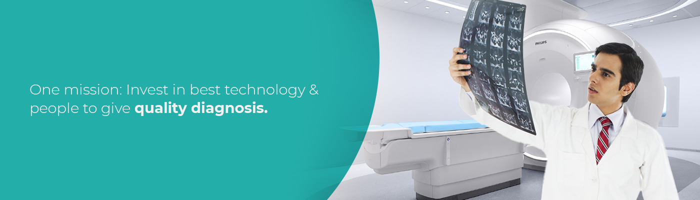
MRI Scan Facility
- Even if you’ve never undergone an MRI scan yourself, you’ve probably heard the term on various medical TV shows or in everyday conversation. In case you aren’t sure what it is, MRI stands for magnetic resonance imaging. It is a diagnostic imaging technique that uses a magnetic field and radio waves to produce detailed images of your body’s internal organs and tissues. An MRI machine can also produce three-dimensional images that may be viewed from several different angles.
- The unit of measurement used to quantify the strength of a magnetic field in an MRI machine is called a Tesla (T). Most MRI scanners operate at a strength of 1.5 Tesla. A 3 Tesla MRI, however, operates at twice the normal strength, providing a greater signal-to-noise ratio, which is a major determinant in generating the highest-quality image.
- The strength of a 3 Tesla MRI yields myriad benefits for radiologists and their patients. Listed below are just a few.
Advantages of Using a 3 Tesla MRI Scanner
- Zero Radiation
- Soft Tissue Demarcation
- Detect flowing blood & vascular malformations
- Detect damages to Myelin Sheath (protective covering certain types of nerves) Superior Imaging
- Free from Beam Hardening & Other Artifacts.
Types of MRI Scans available :-
MRI MRCP
Also Known As MR CHOLANGIO PANCREATOGRAPHY
Magnetic resonance cholangiopancreatography (MRCP) helps visualise the biliary & pancreatic system. The scan includes Pancreas, Pancreatic duct, Bile ducts, Gallbladder and Liver. It is used to evaluate disorders related to health conditions, such as gallstones and pancreatitis.
Test Preparation: 4 Hours of Fasting Required, on the night before the test. Please take 4 tablets of Charcoal and 2 Tablets of Dulcolax: for clearing gases inside the abdomen for better X-Ray results. For Female patients, LMP Date and/or the Latest Pregnancy Test Report is needed.
Reporting TAT: Same Day*
Specialisation(s): Cirrhosis
MRI Brachial Plexus
It is a non-invasive technique that uses a strong magnetic field, radio waves and a computer to make highly detailed images of the brachial plexus, a network of nerves in the shoulder. The scan helps provide a clear structural analysis of the brachial plexus, its intraneural integrity, as well as surrounding structures.
Test Preparation
Male: No Special Preparation. Informed Consent Required.
Female: For Female patients in the reproductive age group, LMP is required.
Reporting TAT: Same Day*
MRI Brain Scan
MRI Brain scan is a painless, safe, and noninvasive test without the use of radioactive tracers/ionizing radiation. It uses a magnetic field and radio waves to create high-quality dimensional images of the brain and brain stem and assists in examining the anatomy of the brain. Brain MRI produces very detailed pictures of the brain.
Test Preparation:
Male/Female: No Special Preparation. Informed Consent Required.
Reporting TAT: Same Day*
Specialisation(s): Neurology.
MRI Brain With Contrast
MRI Scan for the Brain/Head uses a magnetic field and radio waves to create high-quality images of the brain and brainstem. MRI Brain with Contrast allows the MRI machine to see certain parts of the head more easily. There is no use of radioactive tracers/ionizing radiation in the scan which gives a very detailed image of the brain
Test Preparation:
Male/Female: Urea and Creatinine blood test reports are required.
Reporting TAT: Same Day*
MRI Brain with Dynamic CSF Flow Studies
Test Preparation:
Male/Female: No Special Preparation. Informed Consent Required.
Reporting TAT: Same Day*
Specialisation(s): Meningitis, Neurology
MRI lumbosacral (LS) Spine
MRI LS spine is a non-invasive imaging technique that creates high-resolution images of the lumbosacral spine – the bones (vertebrae) in the lower part of the spine. MRI produces high-definition images of the spine area, which are helpful in examining various spine-related diseases or monitoring the progress of the treatment.
Test Preparation:
Male/Female: No Special Preparation. Informed Consent Required.
Reporting TAT: Same Day*
Specialisation(s): Neurology / Orthopaedics
MRI Pelvis
An MRI of the pelvis can help find problems such as tumours in the ovaries, uterus, prostate, rectum and anus. It also can be used to look for an anal fistula (a tube-shaped passage from the anal canal to a hole in the skin near the anus) and look for the cause of pelvic pain in women, such as endometriosis.
Test Preparation:
Female: For Female patients in the reproductive age group, LMP is required.
Reporting TAT: Same Day*
MRI Tongue
The anatomy of the tongue is well demonstrated by magnetic resonance imaging (MRI). On axial T1-weighted images, fat with high signal intensity can be seen between the muscles of intermediate signal intensity. Genioglossus is the largest of all the tongue muscles and forms the bulk of the tongue.
MRI VENOGRAPHY (MRV)
A diagnostic procedure that uses a combination of a large magnet, radio frequencies and a computer to produce detailed images of organs and structures within the body. An MRV uses magnetic resonance technology and intravenous (IV) contrast dye to visualise the veins.
Test Preparation:
Male/Female: No Special Preparation. Informed Consent Required.
Reporting TAT: Same Day*
MRI Whole Spine Screening
A whole spine MRI scan is a non-invasive diagnostic procedure. It uses large equipment capable of creating magnetic fields and radio waves to produce an accurate and clear image of your spine. In addition, the scan image also includes your spinal cord, discs, muscles and other soft tissues in the area.
Test Preparation:
Male/Female: Urea & Creatinine blood test reports are required.
Reporting TAT: Same Day*
MRI Brain with Epilepsy Protocol
It is a painless and non-invasive test that uses a strong magnetic field, radio waves and a computer to make highly detailed images of the brain. The scan identifies areas of brain lesions and blood vessel abnormalities. A contrast agent helps provide a clearer image of the area.
Test Preparation:
Male/Female: Urea & Creatinine blood test reports are required.
Reporting TAT: Same Day*
MRI Brain with Spectroscopy Study
Magnetic resonance (MR) Spectroscopy is a noninvasive diagnostic test for measuring biochemical changes in the brain, especially the presence of tumors.
Test Preparation:
Male/Female: Urea & Creatinine blood test reports are required.
Reporting TAT: Same Day*
MRI Brain with Stroke Protocol
MRI protocol for stroke assessment is a group of MRI sequences put together to best approach brain ischemia.
Test Preparation:
Male/Female: No Special Preparation. Informed Consent Required.
Reporting TAT: Same Day*
MRI Cervical Spine
MRI Cervical spine detects a variety of conditions in the neck and upper back area, including problems with the soft tissues within the spinal column, such as the spinal cord, nerves and disks.
Test Preparation:
Male/Female: No Special Preparation. Informed Consent Required.
Reporting TAT: Same Day*
MRI Dorsal-Lumbar (DL) Spine
MRI Dorsal-Spine is used to see the middle part of the spine to detect various disorders such as congenital deformities of the dorsal spine, diseases of intervertebral discs, nerve compression, and infections of the spine including tumours in the area.
Test Preparation:
Male/Female: No Special Preparation. Informed Consent Required.
Reporting TAT: Same Day*
MRI Fistulogram
Magnetic Resonance Imaging (MRI) for Fistulogram is an imaging technique to create an image of the sphincter muscles, the anal canal, and its surrounding soft tissues. A fistula is an abnormal connection or passageway that connects two organs or vessels that usually do not connect.
Test Preparation:
Male: No Special Preparation. Informed Consent Required.
Female: for patients in the reproductive age group, LMP is required.
Reporting TAT: Same Day*
MRI Knee (Left / Right)
MRI Scan of the Knee creates high-resolution images of the knee joint and the surrounding soft tissues. A knee MRI helps the doctor visualise the anatomy of the knee and find out the cause of pain, weakness or inflammation. There is no use of radioactive tracers/ionizing radiation in the scan.
Test Preparation:
Male/Female: No Special Preparation. Informed Consent Required.
Reporting TAT: Same Day*
MRI Liver For Fat Quantification
It is a non-invasive imaging technique that enables the detection & quantification of liver fat content.
Test Preparation:
Male: No Special Preparation. Informed Consent Required.
Female: For Female patients in the reproductive age group, LMP is required.
Reporting TAT: Same Day*
MRI Lumbar Plexus
Magnetic resonance imaging is an invaluable tool for the evaluation of the lumbosacral plexus due to its anatomic detail and sensitivity to pathologic changes. It can identify the cause of disability, indicate prognosis for improvement, and be a tool for the delivery of interventions.
Test Preparation:
Male: No Special Preparation. Informed Consent Required.
Female: For Female patients in the reproductive age group, LMP is required. Otherwise, a Urine Pregnancy Test is needed.
Reporting TAT: Same Day*
MRI Mammography (also known as MRI Breast)
The scan creates high-resolution images of the inside of the breast. A contrast agent is used for better visualisation of the internal structures. It allows the doctor to screen for breast cancer in women at increased risk, staging of known cancer, and evaluating the response to chemotherapy.
Test Preparation:
Female: Urea and Creatinine blood test reports are required.
Reporting TAT: Same Day*
MRI Prostate
MRI provides information on how water molecules and blood flow through the prostate. This helps determine whether cancer is present and, if so, whether it is aggressive and if it has spread. Sometimes, an MRI of the prostate is needed to evaluate other prostate issues, including infection or abscess.
Test Preparation:
Male: Urea and Creatinine blood test reports are required.
Reporting TAT: Same Day*
Specialisation(s): Prostate
Mri of Upper / Lower Abdomen
The MRI scan creates images of the different organs in the abdomen, such as the bladder, uterus, ovaries and prostate. The images can provide greater clarity on the presence of, for example, cysts, tumours or inflammation.
MRI Whole-Body Diffusion
Whole-body diffusion-weighted magnetic resonance imaging (WB-DWI-MRI) is an emerging tool that has an increasing role in the diagnosis of metastasis and lymphoma. This is a longitudinal study in actual clinical settings designed to assess WB-DWI-MRI in the detection of tumour spread.