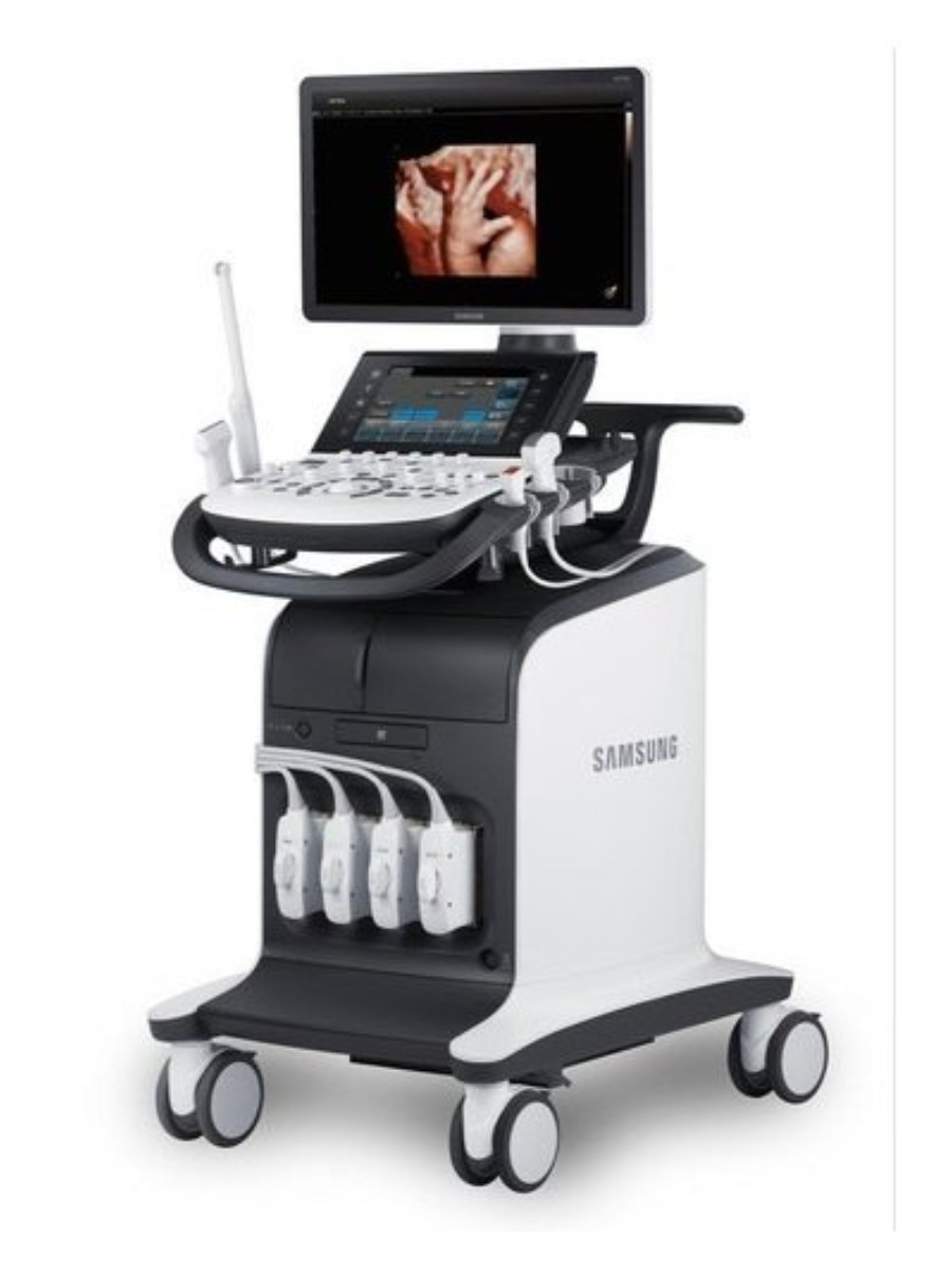
Ultrasonography
Also known as ultrasound or sonography, is a non-invasive imaging procedure that employs sound waves to create visual representations of organs, tissues, and various internal structures within the body. By utilizing ultrasound, healthcare professionals can gain insight into the body without the need for surgical intervention. The resulting images from ultrasound are commonly referred to as sonograms.
Ultrasound has various diagnostic applications & its primary usage are:
- Monitoring the health and growth of developing fetuses during pregnancy. This type of ultrasound, also known as prenatal, fetal, or obstetrical ultrasound, can visualise the baby’s growth, and screen for congenital anomalies (birth-related abnormalities).
- Diagnosing various medical conditions by investigating – organs, glands, blood vessels, etc.
- Assisting in image-guided interventional procedures like FNAC, Biopsy, and Pigtail Catheterization. Ultrasound can precisely locate abnormal areas and guide the needle to the specific location for sample collection.

There are different types of ultrasounds –
- Color Doppler
- Fetal Echo
- USG for Varicose Veins
- Follicular Study
- Pregnancy Sonography
- USG for Deep Vein Thrombosis (DVT)
- Musculoskeletal (MSK)
- Arterial / Veinous Doppler
- Thyroid Scan
- USG of Gyanecological Disorders
- Fibro scan (Liver Stiffness)
- USG for Solid Tumor Stratification
Types of Ultrasound Scans available :-
USG breasts is a safe and painless imaging technique that uses sound waves to create pictures of the inside of the breasts. USG Breasts are safe to use during pregnancy as it does not use any radiation.
Test Preparation: No Special Preparation. Informed Consent Required.
Reporting TAT: Same Day*
USG Foetal Well-Being, also called Obstetric ultrasound, is used to analyze the growth and development of the fetus. It is an imaging process that produces pictures of a fetus within a pregnant woman. This screening method is recommended to begin as early as 7-10 weeks of pregnancy.
Test Preparation: Prescription is mandatory for female patients with a doctor’s sign, stamp, and DMC/HMC number; as per PC-PNDT Act. Please carry your valid Photo ID, such as your Aadhar Card. Past Reports, History and Informed Signed Consent are required.
Reporting TAT: Same Day*
USG KUB test is ultrasonography (USG) or ultrasound of the lower abdomen which assesses the condition of your kidneys, ureters, and urinary bladder (KUB). In the case of males, seminal vesicles and prostate glands are also included in this test.
A level II ultrasound, also known as a fetal anatomical survey, gives detailed information about your baby’s growth. The scan delivers a clear image of the baby’s heart, stomach, brain, and kidneys. It is a painless and safe test usually done during the second trimester (18 to 22 weeks). The test can show whether the pregnant woman has multiple fetuses. It is helpful in the management of twin pregnancies and the complications associated with it.
Test Preparation: Prescription is mandatory for female patients with a doctor’s sign, stamp, and DMC/HMC number; as per PC-PNDT Act. Please carry your valid Photo ID, such as your Aadhar Card. Past Reports, History and Informed Signed Consent are required.
Reporting TAT: Same Day*
It is a safe and painless test that uses sound waves to deliver images of tendons, ligaments, nerves, muscles, and joints throughout the body. Your doctor may advise this test to find out the cause of symptoms such as muscle tears, tendon tears, trapped nerves, sprains, strains, arthritis, masses such as tumours or cysts, and other musculoskeletal conditions.
Test Preparation: No Special Preparation. Informed Consent Required.
Reporting TAT: Same Day*
Ultrasound of the neck is an imaging procedure that helps in visualizing the blood vessels and glands of the neck. It is used to detect any inflammation, cancerous growth, cyst or nodule in the neck region.
Pediatric head ultrasound is a routine procedure that is used to diagnose conditions related to premature birth and to avoid the development of complications in babies that are born prematurely. This module teaches you how to prepare for and perform an ultrasound examination of the pediatric brain, and to assess the common developmental defects. Including both Learn and Test modes, the online simulator offers three clinical scenarios that test your ability to perform an ultrasound examination of the pediatric brain. Practice the steps of the procedures online as often as you want, until you feel confident.
The scan uses high-frequency sound waves to produce images of superficial organs of small parts (including the neck, thyroid gland, and testes). The scan helps determine possible abnormalities in the thyroid, parathyroid glands, scrotum, testis, and breast. It can also be used to guide fine needle aspirations and biopsies of possible abnormalities.
Test Preparation: No Special Preparation. Informed Consent Required.
Reporting TAT: Same Day*
B-scan ultrasonography (USG) is a simple, noninvasive tool for diagnosing lesions of the posterior segment of the eyeball. Common conditions such as cataracts, vitreous degeneration, retinal detachment, ocular trauma, choroidal melanoma, and retinoblastoma can be accurately evaluated with this modality.
Test Preparation: No Special Preparation. Informed Consent Required.
Reporting TAT: Same Day*
This ultrasound examination is usually carried out vaginally but can be done abdominally at around 7 weeks onwards. It aims to determine the number of fetuses present and whether the pregnancy progresses normally inside the uterus.
Test Preparation: Prescription is mandatory for female patients with a doctor’s sign, stamps, with DMC/HMC number; as per PC-PNDT Act. Please carry your valid Photo ID, such as your Aadhar Card. Past Reports, History and Informed Signed Consent are required.
Reporting TAT: 1 Day(s)*
It is a series of transvaginal ultrasound scans that monitors the entire growth process of a follicle from the start of the menstrual cycle to the release of an egg. It forms a vital part of fertility treatments. It helps doctors find out when the patient will ovulate and the number of mature eggs that will ovulate. It also helps to find out the thickness of the Uterine Lining. Ultrasound monitoring also allows the doctor to determine how a woman responds to fertility medication.
Test Preparation: Prescription is mandatory for female patients with a doctor’s sign, stamp, and DMC/HMC number; as per PC-PNDT Act. Please carry your valid Photo ID, such as your Aadhar Card. Past Reports, History and Informed Signed Consent are required.
Reporting TAT: Same Day*
The nuchal translucency test measures the nuchal fold thickness. This is an area of tissue at the back of an unborn baby’s neck. Measuring this thickness helps assess the risk for Down syndrome and other genetic problems in the baby.
Scrotal ultrasound is an imaging test that looks at the scrotum. It is the flesh-covered sac that hangs between the legs at the base of the penis and contains the testicles. The testicles are the male reproductive organs that produce sperm and the hormone testosterone.
A transrectal ultrasound (TRU) used high-frequency sound waves to create images of the organs in the pelvis. The scan helps visualize body parts such as the rectum, prostate (in men), ovaries (in women), and pelvic lymph glands. Your doctor may advise this test to assess the condition of the prostate gland, diagnose certain cancers, pinpoint the location of a tumour in the anus or rectum, check the female pelvic region and examine the size of a tumour.
Test Preparation: No Special Preparation. Informed Consent Required.
Reporting TAT: Same Day*
A Whole Abdomen Ultrasound produces images of internal structures in the abdomen like the appendix, intestines, liver, gall bladder, pancreas, spleen, kidneys, or bladder. Ultrasound Whole Abdomen is used to assess the organs and structures within the abdominal cavity. Ultrasound scans are safe and painless and do not involve ionizing radiation.
Test Preparation: Male: Fasting for 6-8 Hours Female: Prescription is mandatory for female patients with a doctor’s sign, and stamp, with DMC/HMC number; as per PC-PNDT Act. Please carry your valid Photo ID, such as Aadhar Card. Past Reports, History and Informed Signed Consent are required.
Reporting TAT: Same Day*
It is a safe and painless scan that is done on a 20th-week pregnant woman. It helps determine whether or not the embryo in the mother’s womb is developing properly. The scan provides a clear picture of the baby’s heart, stomach, brain, umbilical cord, kidneys, and more. During the test, birth defects can also be detected.
Test Preparation: Prescription is mandatory for female patients with doctors’ signs, stamps, and DMC/HMC numbers; as per PC-PNDT Act. Please carry your valid Photo ID, such as your Aadhar Card. Past Reports, History and Informed Signed Consent are required.
Reporting TAT: Same Day*

