Importance of USG Level 2 / Tiffa / Anamoly during pregnancy

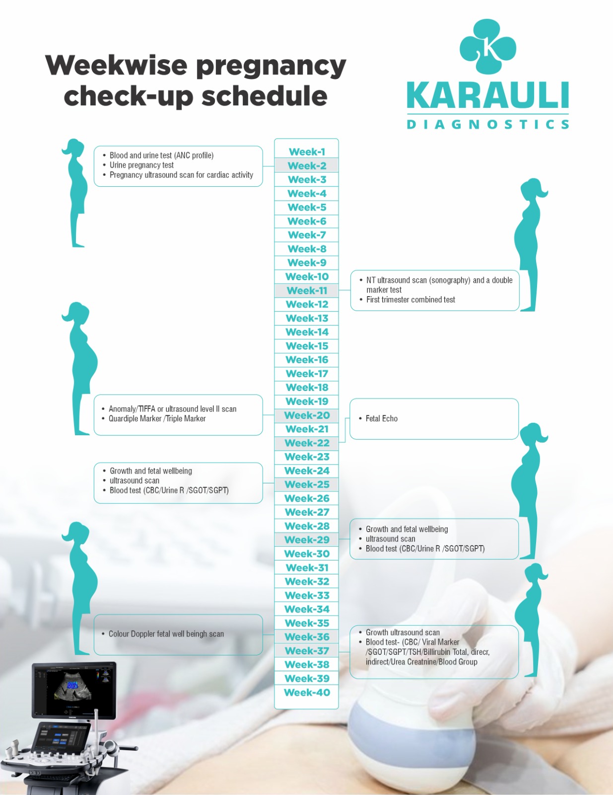
Level 2 ultrasound is a medical imaging procedure that is performed during pregnancy to examine the developing fetus in detail. This type of ultrasound is also called a targeted or detailed ultrasound and is typically conducted between 18 and 22 weeks of gestation (the period of time that a fetus develops inside its mother’s body). It is a more comprehensive examination than the standard ultrasound that is usually performed earlier in the pregnancy.
During a Level 2 ultrasound, a trained ultrasound technician or sonographer will use high-frequency sound waves to create detailed images of the developing fetus. The exam is typically performed abdominally, although in some cases, a transvaginal ultrasound may be used.
The Level 2 ultrasound allows healthcare providers to assess the fetus’s anatomy and development in detail, including the brain, spine, heart, lungs, kidneys, stomach, and other internal organs. The exam can also provide information about the fetus’s growth, the amount of amniotic fluid, and the location of the placenta.
The Level 2 ultrasound is highly recommended during pregnancy for women, fetal abnormalities or who have had abnormal results on other prenatal tests. It can also be recommended for women who have a history of pregnancy complications or who are carrying multiples.
If the ultrasound identifies any potential concerns or abnormalities, further testing or follow-up may be recommended to ensure the best possible outcome for the pregnancy.

The number of ultrasounds to perform during pregnancy varies depending on the healthcare provider’s recommendation and the mother’s medical history. Typically, pregnant women have at least one ultrasound during the first trimester, known as the dating ultrasound, to confirm the due date, rule out ectopic pregnancy, and detect multiple pregnancies.
Additionally, most women have a second ultrasound during the second trimester, known as the anomaly scan, to check the fetus’s growth and development and detect any potential abnormalities. However, some women may require additional ultrasounds if there are concerns about their pregnancy or medical conditions that need monitoring. It is best to consult with your healthcare provider regarding the recommended number of ultrasounds for your pregnancy.
- Early Pregnancy (6 to 8 weeks)
- Confirm viability
- Establish gestational age
- Determine the number of fetus’s
- Determine if multiples share a placenta and amniotic sac
- Nuchal translucency ultrasound (10–13 Weeks)
NT screening, which measures the space behind a fetus’s neck, can detect Down syndrome and other congenital disabilities in the heart, abdomen, and skeleton. Along with an abdominal ultrasound, this screening also involves measuring hormones and proteins through a blood test.
- Anomaly / Tiff a / Level 2 scan (18–22 Weeks)
The anomaly scan, also known as a level II ultrasound, is a detailed pregnancy ultrasound that usually occurs between weeks 18 and 22 of the second trimester. It is the most comprehensive check-up a fetus receives before birth. During the scan, healthcare providers examine the fetus’s brain, heart, kidneys, and liver for abnormalities, and count their fingers and toes. They also check the placenta and measure the amniotic fluid level.
- Special situation ultrasounds
If you have certain medical conditions that require particular monitoring or if you are undergoing a procedure that requires ultrasound guidance, your healthcare provider may recommend these ultrasounds.
Doppler ultrasound:-
Fetal Doppler ultrasound may be recommended by your healthcare provider in the following situations:
– If you have diabetes
– If you have high blood pressure
– If you have heart or kidney problems
– If the placenta fails to develop correctly
– If there are suspected fetal growth problems
Ultrasound is a safe prenatal test performed by healthcare providers, as it uses sound waves instead of radiation like X-rays. Ultrasound has been utilized for over three decades and has not shown any significant risks. Although it is efficient in detecting problems in a healthy pregnancy, it may not be able to detect all birth defects. In some cases, a routine ultrasound can create a false alarm by suggesting a birth defect when there isn’t one, causing unnecessary worry for parents, even though follow-up tests often indicate the baby is healthy.
Common Kinds:-
- Whole-Abdomen ultrasound.
- Transvaginal ultrasound.
In special cases:-
- Doppler ultrasound
- 3-D ultrasound
- 4-D ultrasound
Ultrasound is beneficial in pregnancy because it enables healthcare providers to monitor the growth and development of the fetus. It can help detect potential problems such as ectopic pregnancy, multiple pregnancies, and fetal abnormalities. It is also used to confirm the due date and the position of the placenta.
Furthermore, unlike X-rays, ultrasound uses sound waves, which are safe for both the mother and the baby.
Ultrasound, also known as sonogram, is a prenatal test commonly offered to pregnant women. It employs sound waves to produce an image of the fetus in the uterus. This test enables healthcare providers to monitor the baby’s growth and health.
Ultrasounds use a tool called a transducer that uses sound waves to create pictures of your baby on a computer.

No comment